


Cartilage Preservation

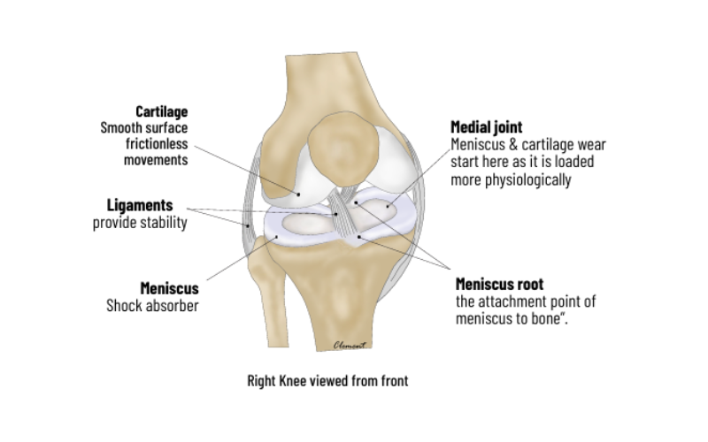

The Knee Joint
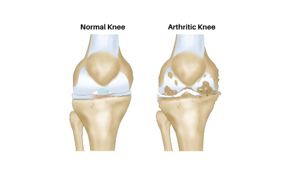
The knee joint is formed by Femur (thigh bone), tibia (leg bone) and patella Damage to the joint is called arthritis and is caused by Obesity, Meniscus tears, Malalignment (“bow-leg”) deformity overloading one compartment, instability (ligament tears) and rheumatological disorders.
Arthritis- Non operative treatment
- Weight loss
- Muscle strengthening exercises
- Acitivity and Lifestyle modification
- Injections like cortisone and
- hyaluronate give short term pain relief.
- PRP and stem cells injections are
being promoted, but are inferior to
cortisone in pain relief and long term
benefits are still questioned. - Unloading braces can off-load the
painful part of knee. - Footwear modifications




Impingement
Impingement refers to abnormal rubbing of the tendons against the shoulder blade end ( acromion). This can occur due to weak cuff muscles (age) that are unable to keep the ball contained or due to bony prominences of acromion irritating the tendon.
Symptoms : Pain with overhead activity ( painful arc), reaching back and pain on sleeping on the same side.
Treatment : Initial conservative – physio and sub acromial injections.
Resistant cases : Arthroscopy, debridement, sub acromial bursectomy ( removal of the inflamed bursa) and
acromioplasty (smoothening of the undersurface of the acromion bone to reduce the
irritation on the tendon.
Treatment
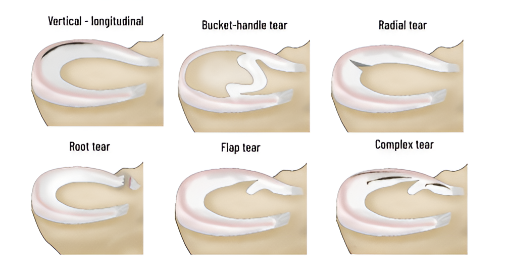
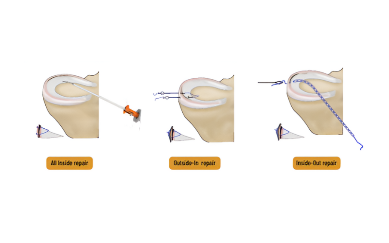
The meniscus has very poor blood supply- only in its rim. Tears in this healing zone are repaired with a variety of techniques. Tears in the non-healing zone and degenerated tears in elderly are treated




Meniscus roott ears
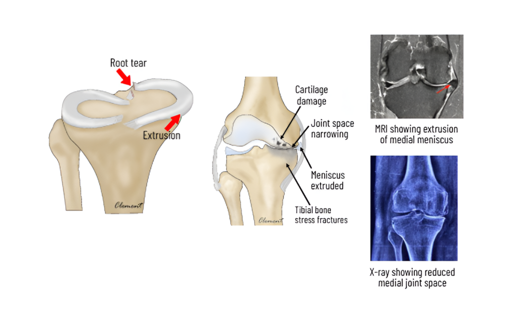
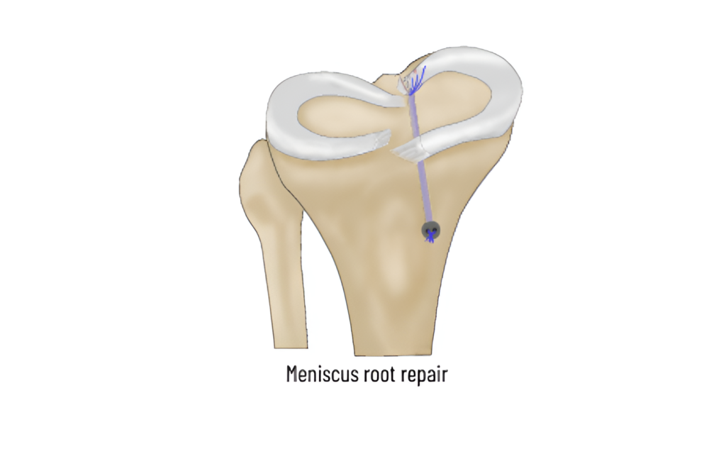
Root tear refers to detachment of meniscus from its bony attachment. It is a devastating injury as the meniscus is pushed out of the joint and shock absorption is completely lost . This results in overloading of the inner compartment leading to accelerated wear and tear ( arthritis)
Treatment
Root repair is done using a keyhole procedure called arthroscopy. Two to three strong sutures are placed in the torn end and brought out through a tunnel in the bone. The sutures are fixed onto the tibia bone using titanium buttons. This root repair restores the protective function of the meniscus. Symptoms History of a minor twisting injury or strain with a popping sensation, followed by pain on the inner side of the knee—worsening at night while lying down, when the knees touch each other, and during turning positions in bed—may lead to bow-leg deformity and limping in the later stages.




Malalignment
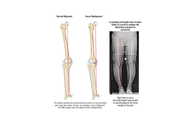
Faulty knee alignment (bow-leg or knock-knee) overloads one compartment, resulting in cartilage injury, meniscus tears and joint space narrowing. As the joint space reduces, the alignment also worsens leading to a vicious cycle of joint destruction and deformity. Fortunately correction of the alignment at an early stage can restore knee function and postpone joint replacement
HTO-High Tibial Osteotomy
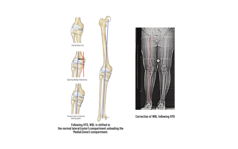
HTO is a procedure to correct the alignment and redistribute the load to the non-involved compartment. Arthroscopy is performed to assess the status of the joint and to treat meniscal tears, loose bodies, and cartilage tears. Based on preoperative planning and using precision instrumentation, a partial bone cut (osteotomy) is made in the upper tibia and is slowly opened to the desired level to shift the load to the normal compartment. The osteotomy is fixed with strong PEEK or titanium plates to allow early movements and weight-bearing. HTO also protects procedures like meniscus repair and cartilage repair by unloading the joint. HTO can postpone the need for knee replacement for many years.




Cartilage injury
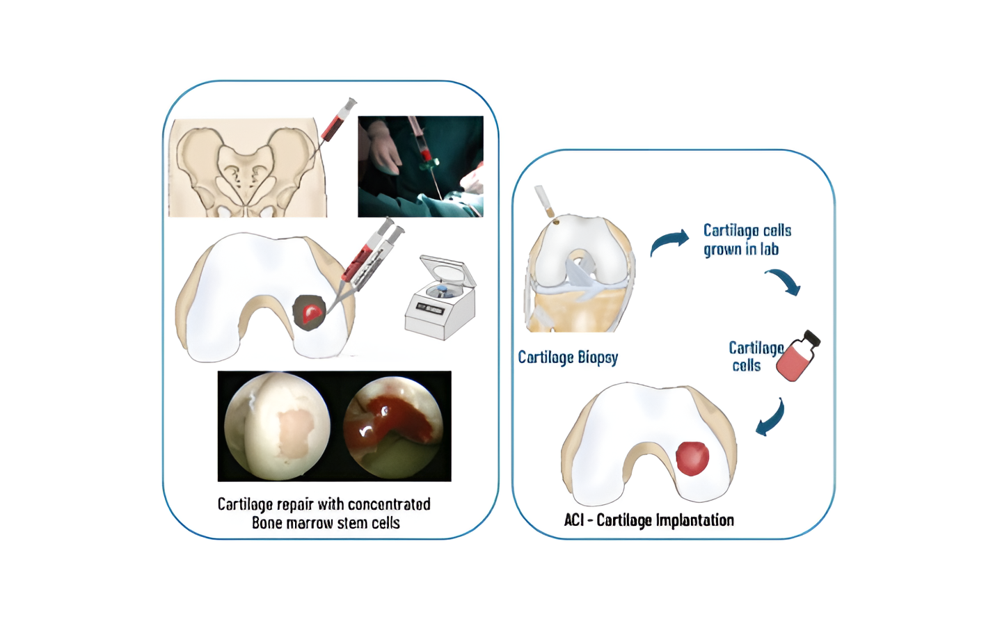
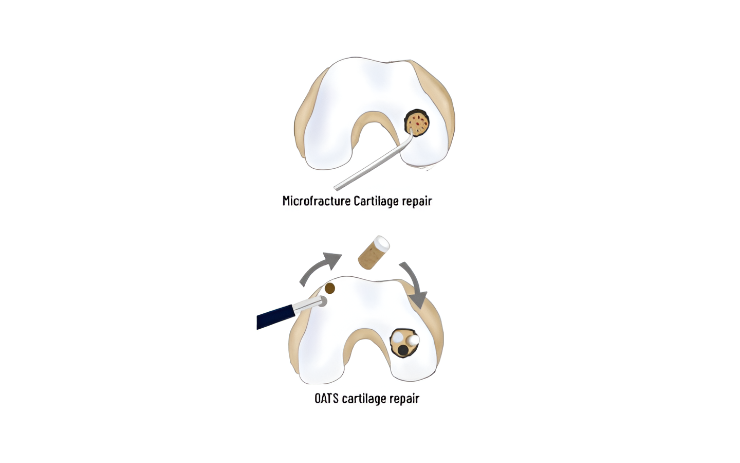
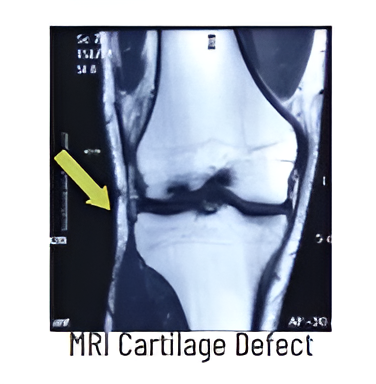
Knee joint surface is covered by Hyaline Cartilage, which is responsible for smooth and frictionless movements. When injured it does not heal due to lack of blood supply. Repair techniques include – MICROFRACTURE (bone marrow stimulation with drilling) suitable for small lesions, OATS, in which cylindrical plugs of cartilage from non-weight bearing areas are transferred to thedefects and Cell based techniques in which concentrated stem cells from bone marrow (BMAC) or patient’s own cartilage cells (ACI) grown in a lab are used to fill the defect.
Treatment
Focal cartilage defects, are treated with variety of techniques. Small defects are treated with Microfracture- marrow stimulating techniques. Large defects are treated with cartilage plug transfers (OATS, mosaicplasty), ACI (cartilage cell culture & repair) and single stage BMA cartilage. Diffuse cartilage wear may need HTO in early stage or TKR in advanced stage

